Welcome to Braces Guide!
After the information gathering has been accomplished and an orthodontist is selected, the treatment process begins. The start of the braces process generally begins after the patient and parents have met with the orthodontist for an initial exam, and have decided that treatment is necessary. The next step is typically an appointment for orthodontic records.
Orthodontic Records for Braces (Video)
The records are a vitally important part of the treatment process. They will provide the orthodontist with much needed information about the patient to make an accurate diagnosis and develop an appropriate treatment plan. Orthodontic records are usually comprised of the following: A clinical exam, Intra and extraoral photographs, Panoramic X-ray, Cephalometric X-ray, sometimes a Cone Beam CT (CBCT), and Impressions (models) of the teeth and bite.
Clinical Exam and Photos
The clinical exam is an extension of the examination performed at the initial exam. A more thorough review of the dental, soft tissue, and jaw problems are recorded to form the basis of the diagnosis. Pictures are taken of the patient outside the mouth (facial and profile photos), as well as inside the mouth to record the upper/lower teeth, as well as record how the patient is biting.
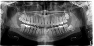
Panoramic X-ray
A panoramic x-ray is crucial to fully visualize the entire upper and lower teeth and jaws (shown below). The x-ray gives the orthodontist information about the jaw bone, roots, jaw joint, as well as evaluating for the presence of extra teeth, impacted teeth, or missing teeth.
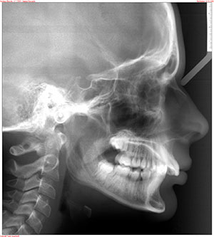
Cephalometric X-ray
A cephalometric x-ray is another x-ray taken during the records appointment. This x-ray provides important information regarding the position of the jaws and front teeth, as well as providing a baseline starting point to monitor growth in younger patients.
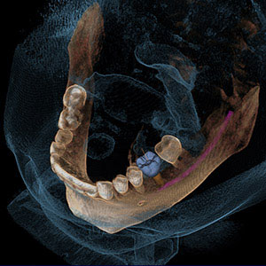
Cone Beam CT (CBCT)
A newer technology in orthodontic imaging is starting to gain more popularity. Cone Beam CT, or CBCT, provides much more information that can be helpful in diagnosis and treatment planning for braces. The CBCT provides a 3-dimensional view of the teeth and jaws, so it can provide spatial information, which is especially important in Cleft Palate patients, or patients that may have impacted teeth, for example. One CBCT exposure can produce many different views for the orthodontist, including the standard panoramic and cephalometric views.
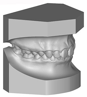
Models
Models of the teeth are made by taking impressions or “molds” of the upper and lower teeth. The models of the teeth are vital for the orthodontist during the treatment planning process. An impression of the teeth is made by placing a soft alginate material in a tray, placing the tray in the mouth, and then waiting about one minute while the material sets up. Alginate feels very similar to pudding or mashed potatoes when it is first placed in the mouth. Once it starts to set up, it has a more rubbery consistency.

After it is removed from the mouth, plaster is poured into the impression. The plaster model is then trimmed to show how the teeth bite together. Some orthodontists have the plaster models scanned digitally, which provides an accurate reproduction of the model. This technology also allows accurate measurements, visualization, and storage.
Digital scanning
A more recent technology has emerged that allows an orthodontist to obtain the 3D image of the teeth and bite through a digital scanning process. This process involves using a capture wand, moving it slowly around the teeth and capturing images from all angles of the teeth. The software then stiches these images together to form the digital model, thereby eliminating the need for the traditional physical impression.
